
Most peptide loss comes down to three overlapping classes of degradation pathway: chemical modification (hydrolysis, deamidation, oxidation), enzymatic cleavage (endo-/exopeptidases, TOP, DPP4) and physical loss (aggregation, adsorption, precipitation). Before investigating anything else, run an LC-MS peptide map of the sample, standardise handling across freeze-thaw cycles and add a protease-inhibited control. Which pathway dominates depends strongly on sequence motifs (Asn, Asp, Met content) and solution conditions such as pH and ionic strength.
TL;DR:
- Peptide degradation often rests on specific chemical modifications such as oxidation and deamidation, which can be identified from characteristic mass shifts.
- Enzymatic cleavage is typically faster than chemical hydrolysis and depends strongly on the peptide’s sequence motifs, such as Pro, Gly or residues exposed to metal ions or light.
- Physical loss through aggregation, adsorption or precipitation can mimic degradation, but can be reduced with gentle handling, low-binding vessels and pH screens.
- Mass spectrometry, particularly LC-MS/MS peptide mapping, is indispensable for diagnosing the exact degradation pathway before specific stabilisation strategies are applied.
- Using COA-verified starting peptides secures correct baseline conditions, since pre-existing degradation products can obscure the actual stability results.
Table of contents
- Peptide degradation pathways: a working taxonomy
- Chemical degradation mechanisms behind peptide bond hydrolysis and oxidation
- How enzymes drive peptide catabolism and intracellular clearance
- Physical loss: aggregation, adsorption and clearance mechanisms
- Detecting peptide degradation with mass spectrometry and peptide mapping
- Stabilisation strategies that target specific degradation pathways
- Designing stability experiments that isolate the right pathway
- Modelling peptide turnover rates and half-life across tissues
- Building a stability checklist that actually works
- Why COA-verified peptides remove a variable from every stability study
- Sources
- FAQ
Peptide degradation pathways: a working taxonomy
Peptide catabolism breaks down into three mechanistic categories, and knowing which one you are fighting changes the entire experimental strategy. Chemical degradation happens without any enzyme at all. It is driven by the intrinsic reactivity of the peptide backbone and side chains under the chosen pH, temperature and buffer conditions. Enzymatic degradation requires a catalyst. Proteases and peptidases recognise specific bonds and cleave them, often at rates orders of magnitude above the equivalent uncatalysed chemistry. Physical degradation is not chemistry at all. Here the peptide drops out of solution, sticks to a tube wall or unfolds into an aggregate that no longer behaves like the intended molecule.
Sequence plays a bigger role in troubleshooting a stability failure than most researchers assume. A peptide with an Asn-Gly or Asp-Gly motif is a near-guaranteed hotspot for deamidation or isomerisation, regardless of everything else in the formulation. Methionine and tryptophan residues are exposed to oxidative attack, particularly under light or in the presence of trace metal contamination. Short N-terminal sequences with Pro or Gly in the second position are classic diketopiperazine risks. Structure matters too. A flexible, solvent-exposed loop degrades faster than the same sequence buried in a folded domain, because both backbone hydrolysis and side-chain oxidation depend on solvent accessibility.
The fastest triage step in any peptide lab is to assign an observed mass shift to a mechanism before touching a single reagent:
- +16 Da on the intact mass usually means oxidation, most often at Met, Cys, Trp or Tyr.
- +1 Da (about +0.984 Da) signals deamidation, which converts Asn or Gln into Asp or isoAsp.
- −18 Da points to cyclisation or dehydration, often linked to the formation of a succinimide intermediate.
- Discrete mass losses corresponding to particular residues indicate proteolytic truncation, by either endopeptidase or exopeptidase activity.
- No mass change but a declining peak area usually means physical loss: adsorption, aggregation or precipitation rather than a chemical conversion.
This assignment is no substitute for full LC-MS/MS peptide mapping, but it tells you where to point the instrument first. Anyone seeing a clean +16 Da shift does not need to run a protease panel. They need a metal chelator and a light-protected storage condition.
Chemical degradation mechanisms behind peptide bond hydrolysis and oxidation
Chemical degradation is the baseline decay rate every peptide is exposed to, even in a sterile, protease-free buffer in the dark. Understanding the triggers of each mechanism is what separates a formulation that survives six months at 4 °C from one that falls apart in a week.
1. Hydrolysis. Cleavage of peptide bonds under neutral or near-neutral conditions is slow compared with enzymatic catalysis, but not negligible over weeks or months. Acid-catalysed hydrolysis accelerates sharply below pH 3, while base-catalysed hydrolysis increases above pH 8. The Asp-Pro bond is a well-known outlier. It hydrolyses far faster than other backbone bonds even at moderate pH, because the neighbouring proline ring strains the geometry of the amide bond. Aspartate residues generally accelerate cleavage of the adjacent backbone through neighbouring-group participation, in which the side-chain carboxylate takes part directly in the hydrolysis mechanism.
2. Deamidation and isomerisation. This is arguably the most sequence-predictable degradation pathway in the whole field. Asn and Gln side chains cyclise via a five-membered succinimide intermediate, which then hydrolyses to a mixture of normal Asp and the structurally altered isoAsp, or racemises to D-Asp. The reaction accelerates at alkaline pH and depends strongly on the residue immediately following Asn or Gln. Gly and Ser in that position accelerate succinimide formation, because they offer little steric hindrance to the backbone geometry required for cyclisation. Bulkier residues such as Val or Ile slow it down. This is why two peptides with nearly identical sequences can show completely different deamidation kinetics.
3. Diketopiperazine formation. Short peptides, particularly those with Pro or Gly in position two, are prone to a specific N-terminal cyclisation in which the first two residues fold back and form a six-membered diketopiperazine ring, releasing the rest of the chain. In the mass spectrum this appears as an unexpected truncation product with a mass loss corresponding to the released fragment, which often confuses researchers who assume they are seeing proteolysis. It is a purely chemical reaction that also occurs in enzyme-free buffer systems.
4. Oxidation. Met, Cys, Trp and Tyr are the four residues that absorb most oxidative damage. Met oxidises to methionine sulfoxide with a clean +16 Da shift, and further oxidation to the sulfone adds another +16 Da. Metal-catalysed oxidation, often from trace iron or copper contamination in buffers, generates reactive oxygen species that attack the same residues, and light exposure accelerates the process considerably, particularly for Trp. Peroxide contamination in polysorbate excipients is a well-documented, frequently overlooked source of oxidation in peptide formulations.
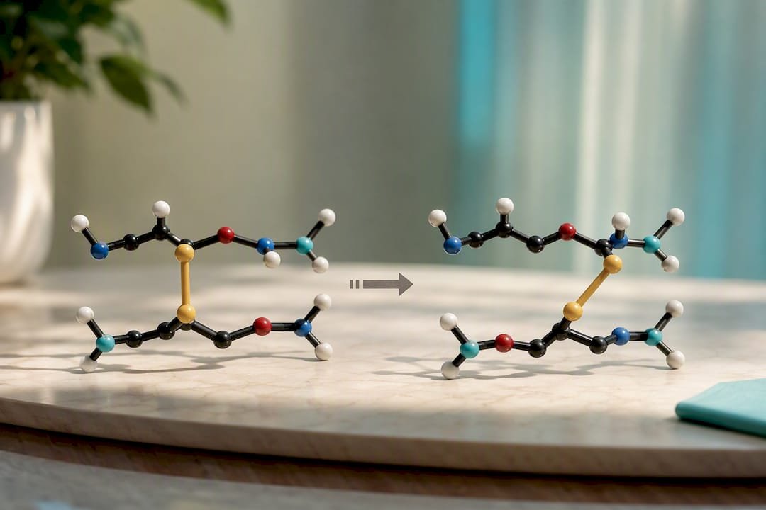
5. β-elimination and disulfide exchange. Under alkaline conditions, Cys and Ser residues can undergo β-elimination, producing a dehydroalanine intermediate that can react further or cross-link. Disulfide-containing peptides carry a separate risk: disulfide scrambling under mildly reducing or alkaline conditions, in which the wrong pair of cysteines re-forms a bond and alters the folded structure without changing the mass.
Pro tip: If a mass loss appears without a shift, do not reach for the protease panel yet. Run a parallel light-stress and metal-chelator condition first — that separates oxidative from proteolytic causes in a single afternoon, before reagents are committed to an enzyme inhibitor screen.
Reviews of therapeutic peptide stability list the same seven chemical pathways as the main drivers of in vivo peptide degradation, and the same chemistry governs how peptides behave in a buffer on the bench.
How enzymes drive peptide catabolism and intracellular clearance
Enzymatic degradation runs faster than chemistry and is specific in ways that reward careful experimental design. Endopeptidases cleave internal bonds and cut a peptide into two or more fragments, based on recognition of a particular residue or motif near the cleavage site. Exopeptidases work from the ends, removing one residue at a time from either the N-terminus (aminopeptidases) or the C-terminus (carboxypeptidases). Research peptides in plasma or tissue homogenates are exposed to both at once, which is why degradation profiles in complex matrices often show a ladder of sequential truncation products rather than a single clean cut.
Thimet oligopeptidase (TOP) deserves particular attention, because it targets peptide length so narrowly. TOP is a metallopeptidase with a strong preference for peptides between 6 and 17 residues, a size range that covers a large share of the research peptides on the market. Inhibition studies show that blocking TOP activity leads to substantial retention of peptides that would otherwise be degraded intracellularly, which means TOP can account for a significant share of degradation in cell extract systems. If the peptide falls into this length window and an unexplained loss shows up in a cell-based assay, TOP inhibition belongs among the first things to test, not in a footnote.
DPP4 (dipeptidyl peptidase 4) works differently and is mainly relevant for peptides with specific N-terminal sequences. It cleaves dipeptides from the N-terminus when a proline or alanine sits in the second position, a motif that is common in many metabolic and incretin-related peptides. This is exactly why GLP-1 analogues and similar peptides are subject to rapid plasma clearance unless this N-terminal weak point is designed around. Beyond DPP4, plasma contains a broad range of proteases, and tissue distribution studies identify liver, kidney and gastrointestinal tissue as particularly high-activity sites of peptide hydrolysis, a factor that matters enormously when choosing an in vitro matrix intended to predict in vivo behaviour.
Inside the cell, degradation runs through entirely different machinery. The ubiquitin-proteasome system tags proteins and larger fragments with ubiquitin chains for proteasomal degradation, while the autophagy-lysosome pathway processes bulkier structures, including aggregates that the proteasome cannot process efficiently. Together these two systems form the primary intracellular clearance mechanism for peptide and protein fragments, and they operate on a completely different timescale and with different substrate specificity from plasma proteases.
Designing an experiment that confirms which enzyme system is responsible for a given loss comes down to a few concrete steps:
- Run a broad-spectrum protease inhibitor cocktail alongside an untreated control in the same matrix.
- Use protease-depleted or heat-inactivated matrix as a negative control, to isolate chemical from enzymatic degradation.
- Test recombinant TOP or DPP4 directly against the peptide when sequence or length matches their known substrate profiles.
- Compare plain plasma incubation with cell lysate or tissue homogenate, since intracellular and extracellular enzyme populations differ considerably.
The substrate window of TOP (6 to 17 residues) is narrow enough that a quick check of sequence length shows within seconds whether it belongs on the inhibitor shortlist.
Physical loss: aggregation, adsorption and clearance mechanisms
Not every disappearing peptide has been chemically altered or enzymatically cleaved. A surprisingly large share of the “degradation” observed in the lab is actually physical loss, and it is often the easiest problem to fix once correctly identified.
Aggregation occurs when peptide concentration, agitation or the absence of a stabilising excipient pushes molecules together into insoluble or soluble oligomers. Highly concentrated formulations are particularly susceptible, and even mild shaking during shipping or handling can trigger aggregate formation in a peptide that looked perfectly stable sitting still in the vial.
Adsorption to glass and plastic surfaces is a chronic, underestimated source of apparent concentration loss, especially at low peptide concentrations, where a considerable part of the total material can stick to the wall of a standard polypropylene tube. Switching to low-binding consumables or adding a small amount of surfactant such as polysorbate 20 to the buffer usually solves the problem without touching the peptide’s chemistry at all.
Precipitation shows up when pH approaches the peptide’s isoelectric point, or when changes in ionic strength abruptly alter solubility. A solubility screen across a pH range, run before a final buffer is fixed, catches this before it becomes a mysterious loss during a longer stability study.
In vivo, renal clearance is the dominant physical route of elimination for small peptides. Molecules below the renal filtration threshold, around 60 kDa, but effectively far lower for typical research peptides, are filtered out by the kidney within minutes to hours, unless a conjugation or formulation strategy extends their circulation time.
- Reduce agitation-driven aggregation with gentle handling protocols and minimised headspace in storage vials.
- Use low-binding tubes and consider a carrier protein or surfactant for dilute, adsorption-prone samples.
- Screen solubility across pH and ionic strength before fixing a working buffer.
- Account for renal clearance explicitly when interpreting in vivo half-life data, since it can dominate any chemical or enzymatic contribution for small peptides.
Detecting peptide degradation with mass spectrometry and peptide mapping
LC-MS/MS peptide mapping sits at the centre of any serious degradation investigation, and for good reason. It combines intact mass measurement, which quickly shows whether anything has changed at all, with MS/MS fragmentation, which pinpoints exactly where in the sequence a modification has occurred. Intact mass screening is the fast first pass. Run at time zero and at the study endpoint, any shift outside instrument error shows that something has happened, even before you know what.
MALDI-TOF offers a faster, lower-resolution alternative for a rapid screen across many samples, useful when triaging a large number of stability conditions before committing MS/MS time to those that actually show change. Capillary electrophoresis adds a complementary dimension of separation, particularly useful for distinguishing charge variants such as isoAsp and Asp, which have identical mass but different electrophoretic mobility.
Reading the mass shifts correctly is the skill that separates a quick diagnosis from weeks of guessing:
- +16 Da almost always means oxidation at Met, Cys, Trp or Tyr; MS/MS determines the residue affected.
- +0.984 Da (rounded to +1 Da on lower-resolution instruments) indicates deamidation, converting Asn or Gln into Asp/isoAsp.
- Discrete losses matching one or more residue masses point to exopeptidase trimming or endopeptidase cleavage.
- −18 Da suggests dehydration or cyclisation, often linked to the formation of a succinimide intermediate en route to isomerisation.
- An unexpected mass gain matching another peptide or protein fragment signals cross-linking, sometimes disulfide-mediated.
None of these signatures should stand alone as a definitive answer. Orthogonal validation matters. An enzymatic digest followed by peptide mapping confirms proteolytic cleavage sites independently of intact mass data. Activity assays confirm whether a chemically modified peptide still performs its function, which matters enormously when evaluating a stabilised analogue. SEC or SDS-PAGE captures aggregation that mass spectrometry alone might miss, since aggregates do not always ionise or fragment the way monomeric peptide does.
Pro tip: The protease-inhibited control and the untreated sample should be run side by side under identical LC-MS/MS conditions. If the degradation pattern is identical in both, it is chemistry, not enzymes, and a week of inhibitor screening has just been saved. This foundational mass-spectrometric approach to peptide metabolism and catabolism remains the field’s central diagnostic tool for exactly that reason.
Stabilisation strategies that target specific degradation pathways
Once you know which pathway is degrading the peptide, the solution usually falls into one of five categories, and choosing the right one saves months of trial and error.
1. Sequence modification. Substituting D-amino acids at protease-susceptible positions can extend half-life dramatically. Documented cases show a D-arginine substitution raising a peptide’s half-life from minutes to hours by disrupting the stereospecific recognition most proteases rely on. The trade-off is real: changing stereochemistry can alter receptor binding, so every modified analogue needs an activity assay in addition to the stability test, not instead of it.
2. Terminal protection. N-acetylation blocks aminopeptidase attack from the N-terminus, and C-terminal amidation blocks carboxypeptidase trimming from the other end. This is often the cheapest and lowest-risk modification available, since it typically preserves the internal sequence responsible for biological activity.
3. Cyclisation and backbone modification. Cyclic peptides resist both hydrolysis and protease access, simply because there is no free terminus for exopeptidases to start from, and because the constrained conformation often hides the backbone from the recognition sites of endopeptidases.
4. PEGylation, lipidation and conjugation. Attaching a polyethylene glycol chain, a lipid tail or a carrier protein increases the hydrodynamic radius beyond the renal filtration threshold, extending circulation time considerably while also physically shielding cleavage sites from protease access.
5. Excipient and container selection. Antioxidants such as methionine or ascorbate scavenge reactive oxygen species before they reach susceptible residues. Metal chelators such as EDTA remove the traces of iron and copper that catalyse oxidative chemistry. Low-binding container closures and validated cold-chain handling protect against adsorption and thermal degradation, which have nothing at all to do with the peptide’s intrinsic chemistry.
Every one of these measures has to be verified before it is trusted. Research on chemical modification and testing is consistent on this point: a peptide that resists degradation but no longer binds its target is not a solution, it is a different problem with the same molecular formula.
Designing stability experiments that isolate the right pathway
A stability assay only yields something useful when matrix, controls and handling protocol are chosen to isolate the variable actually being tested.
Matrix selection should match the research question directly. Plasma incubation reports on stability in the circulation, but says almost nothing about intracellular clearance, since plasma and intracellular peptidase populations differ considerably. Tissue homogenates give a rough approximation of local enzymatic activity in liver, kidney or gut, the sites with the highest activity for peptide hydrolysis. Purified enzyme systems such as recombinant TOP or DPP4 isolate a single mechanism cleanly, which is exactly what you want once a hypothesis is to be confirmed rather than a broad screen run.
Controls decide how interpretable any degradation study is. A protease inhibitor cocktail condition should be run alongside the test sample in identical matrix and with identical timing. A heat-inactivated matrix control confirms the enzymatic contribution independently. Always include a blank matrix control without peptide, to capture background interference in the mass-spectrometric evaluation, and quench samples immediately at each time point rather than letting them stand at room temperature before analysis, which artificially raises apparent degradation rates.
Handling details that seem insignificant often dominate the noise in a degradation data set:
- Use low-binding consumables throughout, not only for the final dilution step.
- Minimise freeze-thaw cycles, since repeated freezing stresses both chemical stability and aggregation propensity.
- Quench enzymatic activity immediately at each sampling time point with a cold stop solution or rapid heat denaturation.
- Store reference standards under exactly the conditions used in stability protocols per ICH Q1A(R2), so that results are comparable across studies.
Pro tip: Full assay conditions, raw peptide maps and the COA of the starting material should be recorded in supplementary documentation, even for internal lab notebooks. When a result cannot be reproduced six months later, that record is the only thing that shows whether the peptide or the protocol changed. This level of documentation mirrors the reporting rigour behind the formal guideline on stability testing for regulated active substances.
Modelling peptide turnover rates and half-life across tissues
Fitting a decay curve to degradation data looks simple until the data refuse to follow a single exponential. Many peptides show multiphasic decay: a fast initial phase dominated by rapid enzymatic cleavage or renal clearance, followed by a slower phase reflecting a more resistant subpopulation or a secondary, slower degradation pathway. First-order kinetics work well for isolated chemical reactions such as deamidation in a simple buffer, but plasma or tissue data often need a two-compartment or biexponential model to be fitted honestly.
The distribution of tissue peptidases changes the whole picture depending on where you measure. A peptide’s apparent half-life in plasma can look deceptively long if you fail to account for liver, kidney and gut tissue having far higher peptidase activity than the blood itself. The narrow substrate window of TOP (6 to 17 residues) illustrates this well: a peptide right in that range can show moderate plasma stability but rapid intracellular turnover once it enters a cell, an effect that plasma-only assays systematically miss.
This is exactly why extrapolating in vitro plasma stability directly to in vivo pharmacokinetics without tissue-level data is risky. A peptide that looks stable for two hours in a plasma tube can still be eliminated rapidly once it distributes into kidney or liver tissue with dense peptidase populations. Tissue-specific enzyme activity should be built into the PK model from the start, rather than treating plasma half-life as a universal proxy for whole-body clearance.
Building a stability checklist that actually works
Start with analytics, not assumptions. Run an intact mass screen at the start and at the endpoint of the study before touching a single formulation variable, then LC-MS/MS peptide mapping on every sample that shows a shift. That one step tells you more than a week of blind buffer optimisation.
Once the degradation pathway is known, fix it specifically. Adjustments to buffer and excipients (antioxidants, chelators, pH adjustment) solve most chemical degradation problems cheaply. Enzymatic loss calls first for inhibitor panels or protease-depleted matrices, and sequence modification only once inhibition has confirmed the mechanism. Escalation to PEGylation, cyclisation or conjugation should wait until simpler solutions miss the stability target, since these carry higher development costs and activity risks.
Reproducibility starts with the starting material. A peptide that arrives already partly degraded distorts every subsequent result, which is why sourcing from a supplier with verifiable, batch-specific documentation (disclosed method, chromatogram, named testing lab) matters as much as assay design.
— Max
Why COA-verified peptides remove a variable from every stability study
A degradation study is only as trustworthy as its starting material. If the peptide arrives with unknown purity, or with degradation products already present from an earlier, poorly controlled synthesis run, every mass shift measured afterwards is contaminated by an unknown baseline.

This distinction matters most in exactly the kind of work this article covers. Deamidation, oxidation and truncation studies all depend on a clean t=0 baseline, and a peptide of unverified purity makes it impossible to separate pre-existing degradation from what is being tested in the lab. Peptelia’s product quality and testing page explains the HPLC verification process behind every COA, and researchers working on neuropeptide stability can review a verified starting sample directly on the Selank product page. Before the next stability run, check the current batch documentation.
Sources
- Characterisation of thimet oligopeptidase and its role in peptide degradation (PubMed record)
- In vivo degradation forms, anti-degradation strategies and clinical applications of therapeutic peptides (PMC review)
- Peptide and protein drugs: studying their metabolism and catabolism by mass spectrometry (methods review)
FAQ
What are the main peptide degradation pathways?
Peptide loss falls into three classes: chemical (hydrolysis, deamidation, oxidation), enzymatic (cleavage by proteases and peptidases) and physical (aggregation, adsorption, precipitation, renal clearance).
How do you tell chemical degradation apart from enzymatic degradation?
Run a protease-inhibited control alongside the test sample in identical matrix and with identical timing; identical degradation in both conditions points to chemistry, while suppressed degradation in the inhibited sample confirms enzymatic involvement.
Which analytical method best detects peptide degradation?
LC-MS/MS peptide mapping is the central method; it combines intact mass screening for rapid detection with MS/MS fragmentation for exact localisation of the modification site, as documented in methodological reviews of mass spectrometry.
Why does thimet oligopeptidase matter for short research peptides?
TOP preferentially targets peptides between 6 and 17 residues, and inhibition studies show it can account for a considerable share of the degradation of peptides in that length range in cell extract systems.
How can researchers reduce uncertainty from starting material quality?
Sourcing peptides with a verifiable, batch-specific certificate of analysis (COA) that discloses the method and names the testing lab establishes a verified purity baseline before any stability testing begins.
Does plasma stability predict in vivo half-life accurately?
Not reliably on its own, since the distribution of tissue peptidases in liver, kidney and gut often produces faster clearance than plasma-only incubation would suggest.