Peptide stability depends on three interacting groups of factors: sequence chemistry (hydrophobicity, aromatic residue content, isoelectric point), physical drivers of aggregation (concentration, interfaces, temperature, agitation), and handling and formulation conditions (buffer, excipients, storage). Sequence-based regression models can now estimate serum half-life directly from the percentage of non-polar residues, the number of Tyr/Trp residues and the pI, while the NIBSC storage recommendations cover the handling side. Run a liability scan of the sequence first, then confirm it with a short forced-degradation stress test and a serum half-life assay, before settling on a formulation.
TL;DR:
- Sequence features such as a high content of non-polar residues and several tyrosines improve serum stability, while the presence of tryptophan and a higher pI tend to accelerate degradation.
- Formulation should target specific weak points; buffers such as acetate or citrate are often better than phosphate once deamidation or hydrolysis risks have been identified.
- Peptides should be stored at −20 °C or colder under dry conditions and split into small aliquots after reconstitution, to minimise damage from freeze-thaw cycles and moisture.
- Surface adsorption and mechanical stress from agitation are common causes of physical instability and aggregation, and can be reduced with low-binding vessels and gentle handling.
- Reproducibility depends heavily on detailed documentation of serum batch, pH, buffer and analytical methods; sourcing from verified suppliers is decisive for consistent starting quality.
Table of contents
- Which sequence-dependent factors affect peptide stability?
- What drives peptide aggregation and physical instability?
- Which chemical degradation pathways affect peptides?
- How do buffers and excipients affect peptide formulation stability?
- How should research peptides be stored and handled?
- How is peptide stability tested and measured?
- Which interventions actually improve peptide stability?
- How to structure a peptide stability study step by step
- What researchers get wrong about reproducibility in stability data
- Where to source verified peptides for stability testing
- Sources
- FAQ
Which sequence-dependent factors affect peptide stability?
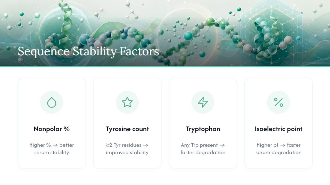
The amino acid sequence sets the upper limit on how stable a peptide can be at all, regardless of how carefully it is stored afterwards. A regression model built from a curated set of published half-lives found that four sequence features explain a substantial part of the variance in serum stability: the percentage of non-polar residues, the number of tyrosine residues, the presence of tryptophan and the isoelectric point. Peptides with a higher proportion of non-polar residues and two or more tyrosines tend to resist proteolytic degradation better than average. Peptides with at least one tryptophan or a higher pI tend to be degraded faster in serum.
This matters because it turns stability prediction into a desk exercise that can be completed before a single milligram is ordered. No wet-lab assay is needed for a first risk assessment. All it takes is a sequence and a spreadsheet.
The liability scan checklist
Before synthesis, or before a candidate goes into expensive serum work, run this five-point scan:
- Calculate the percentage of non-polar residues (Ala, Val, Leu, Ile, Pro, Phe, Met, Trp) across the whole sequence.
- Count tyrosine residues; two or more is a favourable sign for persistence in serum.
- Flag every tryptophan residue, since in the regression model even one is associated with reduced stability.
- Calculate the isoelectric point; sequences with a higher pI showed weaker proteolytic resistance in the same data set.
- Look for Asn, Gln, Met, Trp and free Cys residues, each of which carries its own chemical weak point independent of the aggregate hydrophobicity score.
Statistics note: The multivariable regression model from PMC8301568 was validated against both synthetic peptide libraries and clinically tested peptides, and provides a concrete equation for estimating serum half-life from these four inputs, not just a qualitative risk score.
Residue-specific weak points deserve individual attention, because they are not always visible in an aggregate hydrophobicity or pI calculation. Asparagine and glutamine residues are hotspots for deamidation, particularly when glycine or serine follows in the sequence. Methionine and tryptophan oxidise under even moderate oxidative stress, and free cysteine residues can form unwanted disulfides or thiol-disulfide exchange products during storage. If a target sequence contains one of these residues at a position that is not required for bioactivity, a terminal protecting group (N-terminal acetylation, C-terminal amidation) or a protecting-group strategy can mitigate the exposure without redesigning the whole molecule.
Here lies the trade-off on which much formulation work founders: increasing hydrophobicity to gain proteolytic resistance often raises the aggregation risk at the same time. The same aggregation research that defines the drivers of physical stability also notes that hydrophobic regions are the nuclei of self-association. A sequence change that solves one stability problem can create another, which is why sequence screening and aggregation screening have to happen together rather than one after the other.
What drives peptide aggregation and physical instability?
Aggregation is a kinetic process, not a threshold event. It begins with nucleation, in which a small number of misfolded or self-associating monomers forms a nucleus, and then propagates as further monomers attach to that nucleus. Concentration is by far the biggest lever here: the aggregation rate scales with monomer concentration, often non-linearly, so a formulation that appears stable at 0.1 mg/mL can fail completely at 5 mg/mL. This is one of the most common surprises during scale-up, when a promising result at low concentration no longer holds once the stock is concentrated to give practical dosing volumes.
Interfaces are the second major driver, and they are easily underestimated. Air-liquid interfaces created by shaking, pipetting or the headspace in a partially filled vial can trigger aggregation even at low total concentration. Vessel walls do the same through adsorption: a dilute peptide solution in a standard polystyrene tube can lose a considerable part of its active material to the walls before the assay is even run, which shows up as an apparent stability failure that is in reality only surface loss.
Mechanical and thermal stress aggravate both problems:
- Agitation (vortexing, transport vibration, repeated pipetting) increases exposure at the air-liquid interface and can trigger partial unfolding through shear forces.
- Sonication, sometimes used to dissolve peptides, can paradoxically trigger aggregation if applied for too long or too intensely.
- Freeze-thaw cycles locally concentrate the peptide at the ice-water interface during freezing and create transient zones of high concentration that favour nucleation.
- Elevated temperature accelerates both the chemical degradation pathways and the physical unfolding that precedes aggregation, so a formulation that is stable at 4 °C can behave differently within hours at room temperature.
Pro tip: Before blaming a formulation for poor stability, rule out surface adsorption first. A quick recovery check helps: measure the concentration in a fresh low-binding vial against a standard polypropylene tube after the same incubation time. If recovery differs by more than a few per cent, it is a vessel problem, not necessarily a molecule problem.
Practical control measures follow directly from these mechanisms. Low protein-binding vials reduce adsorption losses to surfaces, which is especially valuable at low working concentrations. Gentle handling, meaning minimal vortexing, slow pipetting and avoiding unnecessary freeze-thaw cycles, protects against mechanically induced nucleation. Surfactants such as polysorbate 20 or 80 can displace the peptide at the interface, though compatibility has to be checked against the specific sequence, since surfactants themselves can sometimes destabilise certain peptides. For monitoring, dynamic light scattering (DLS) captures early aggregation and shifts in size distribution long before visible turbidity appears, while size exclusion chromatography (SEC) quantifies soluble aggregate species and provides a value that can be tracked across formulations.
Which chemical degradation pathways affect peptides?
Four covalent degradation pathways account for the bulk of chemical instability in peptides, and each has its own sequence and condition dependence that should be stress-tested separately rather than lumped together in a generic “stability assay”.
- Hydrolysis. Backbone amide bonds cleave under both acid and base catalysis, with the rate depending strongly on pH and on particular sequence motifs. Asp-Pro bonds are known to be acid-labile, and peptides with this motif can show measurable backbone cleavage over days at even moderately low pH and room temperature.
- Oxidation. Methionine, tryptophan and cysteine residues are the primary oxidation targets; they react with dissolved oxygen, trace metal ions or light-induced reactive species. Controlling headspace oxygen (overlaying with nitrogen or argon), avoiding light exposure and adding antioxidants such as methionine as a scavenger or chelators such as EDTA are standard countermeasures for oxidation-prone sequences.
- Deamidation. Asparagine and glutamine side chains hydrolyse to aspartate and glutamate, with Asn generally reacting faster than Gln and both strongly accelerated at higher pH and elevated temperature. Analytically this shows up as a one-dalton mass shift per deamidation event and as a pI shift, both detectable by LC-MS and isoelectric focusing.
- Isomerisation and racemisation. Harsh conditions, high temperature, extreme pH or prolonged exposure to strong acids or bases during post-synthesis purification can convert L-amino acids into D-amino acids or trigger backbone isomerisation at Asp residues. Neither is usually detected by standard mass spectrometry, since the mass does not change, which is why gentle handling during purification and storage matters more than is often assumed.
Each of these pathways has a different pH and temperature optimum for stability, which is the practical reason why a single instruction to “store at 4 °C” is not enough. A deamidation-prone sequence needs a buffer at lower pH. A sequence prone to acid hydrolysis at an Asp-Pro bond needs the opposite. Knowing which weak point dominates for a given sequence, from the residue scan described earlier, tells you which stress condition to test first.
How do buffers and excipients affect peptide formulation stability?
Buffer choice is not a neutral background decision. Phosphate buffer, one of the most common options in early work, can actually catalyse certain degradation reactions in sensitive sequences. So if a residue scan flags a deamidation or hydrolysis risk, it is worth testing an alternative buffer species such as acetate, citrate or histidine before settling on a formulation. This is a practical precaution, not a fixed rule. Buffer selection should follow the specific weak point of the sequence rather than defaulting to whatever is on the shelf.
Excipients earn their place in a formulation by solving a specific mechanistic problem, and matching excipient to weak point is where formulation science actually pays off:
- Sugars such as trehalose or sucrose act as lyoprotectants and stabilise peptide structure during freeze-drying by forming a glassy matrix that replaces the hydrogen-bonding function of water.
- Surfactants (polysorbate 20/80) reduce interface-driven adsorption and aggregation, which is particularly useful for peptides prone to losses at the air-liquid interface.
- Antioxidants (methionine, ascorbate, EDTA as a metal chelator) target oxidation-prone sequences containing Met, Trp or Cys.
- Bulking agents such as mannitol support cake structure in lyophilised formulations without necessarily protecting against unfolding the way sugars do.
Pro tip: If the sequence scan flags both an oxidation weak point and an aggregation risk, do not combine all the relevant excipients at once. Test them individually first. Antioxidants and surfactants can interact in ways that mask each other’s benefit, and you should know which one is actually doing the work before finalising a multi-component formulation.
The choice of ionic strength and pH involves a real trade-off between solubility and chemical stability. Higher ionic strength can improve the solubility of charged peptides, but it can also promote aggregation by screening the electrostatic repulsion that keeps monomers apart in solution. A pH chosen purely for solubility can sit exactly in the zone of accelerated degradation for a deamidation-prone sequence, so the formulation pH usually has to be a compromise rather than an optimisation of either variable alone.
Lyophilisation brings its own excipient logic. Glass formers have to keep the formulation below its collapse temperature during both freezing and primary drying, otherwise the dried cake loses its structure and rehydrates poorly. Reconstitution should as a rule use the solvent in which the peptide was characterised, added slowly down the vial wall rather than squirted directly onto the cake, and be given a few minutes to dissolve completely before vortexing or agitation.
How should research peptides be stored and handled?
Storage decisions in the first five minutes after a peptide arrives have more influence on its usable lifetime than almost anything that happens afterwards.
- Store lyophilised peptide at −20 °C or colder, with −80 °C preferred for long-term archival storage, based on the NIBSC handling recommendations and manufacturers’ technical documentation.
- Let the vial equilibrate to room temperature in a desiccator before opening, so that atmospheric humidity does not condense on the cold, hygroscopic powder as soon as the seal is broken.
- Aliquot the reconstituted stock into single-use volumes immediately, since repeated freeze-thaw cycles are one of the most common self-inflicted causes of apparent instability in everyday lab work.
- For oxidation-prone sequences containing Met, Trp or Cys, overlay the headspace with an inert gas such as nitrogen or argon before resealing.
- For dilute working solutions, choose low protein-binding polypropylene vials or specially treated glass vials, since standard glass or polystyrene surfaces can adsorb a considerable part of the peptide unnoticed.
- Follow a solvent hierarchy when dissolving: water first, then dilute acetic acid for peptides with basic residues that will not dissolve in pure water, and only if aqueous options fail, a small proportion of DMSO or acetonitrile.
- Use short, low-power sonication if a peptide is hard to dissolve, and filter through a low protein-binding syringe filter before any concentration-sensitive assay.
GenScript’s manufacturer recommendations confirm the same core points, storage at −20 °C or below, minimal moisture exposure and avoidance of repeated freeze-thaw cycles, together with a concrete recommendation for sterile buffers at pH 5 to 6 when short-term storage in solution is unavoidable. The agreement across independent handling recommendations is itself a signal that these are not optional best practices; they are the baseline that separates a usable stock within weeks from a degraded one.
How is peptide stability tested and measured?
Designing a stability study well means choosing assays that map directly onto the weak points the sequence scan has already flagged, rather than running a generic panel and hoping something useful comes out of it.
- Forced degradation. Run parallel stress conditions, oxidative (addition of hydrogen peroxide), pH extremes (buffers at pH 2 and pH 9) and thermal (40 °C to 60 °C for accelerated ageing), each against a room-temperature control across the same time points.
- Serum or plasma incubation. Incubate the peptide in serum or plasma at 37 °C and sample at several time points to measure the proteolytic half-life directly, because that is the endpoint the sequence-based regression models are trying to predict.
- Aggregation assays. Run SEC and DLS in parallel on the same samples used for the degradation tests, since aggregation and chemical degradation often occur together and can confound each other when measured separately.
- Analytical mapping. Use LC-MS to identify and quantify specific degradation products (deamidated species, oxidised variants, hydrolysis fragments) instead of relying on a single aggregate purity figure.
Statistics note: Published protocols for peptide stability testing consistently stress that analytical measures have to be matched to the mechanism, LC-MS for mapping degradation products, SEC and DLS for aggregation and CD spectroscopy where loss of secondary structure is relevant, because a single purity figure can obscure which pathway is actually responsible for the loss.
Matrix effects are the biggest source of misleading results in serum work. Serum origin, lot number and whether the serum was heat-inactivated all shift the observed proteolytic half-life, sometimes considerably, and these are exactly the details that go unmentioned in many published stability comparisons. If a sequence variant is benchmarked against a literature value and the publication names neither serum lot nor heat-inactivation status, the comparison should be treated as indicative, not precise.
Reproducibility is a matter of documentation discipline: buffer composition and pH, enzyme or serum origin and lot, incubation temperature and the analytical limit of detection for the degradation product being tracked. Half-lives without that context cannot be interpreted with confidence by anyone, including your own lab six months later.
Which interventions actually improve peptide stability?
Four categories of intervention come up repeatedly in the literature, each with a different cost-benefit profile depending on how much structural flexibility the target sequence allows.
- Sequence changes. Swapping a labile residue (an exposed Trp, an Asn-Gly deamidation motif) for a more stable alternative works when the residue is not required for bioactivity, and lowering the overall pI can improve persistence in serum based on the regression results discussed earlier.
- Chemical modifications. D-amino acid substitution and N-methylation block protease recognition at specific backbone positions; terminal protecting groups remove exposed charged termini that are common protease attack points; PEGylation and lipidation increase apparent size and can reduce renal clearance and proteolytic exposure, though both can reduce receptor binding affinity if placed too close to the active pharmacophore.
- Conformational constraints. Cyclisation and stapling lock a peptide into a defined secondary structure and physically shield the backbone amide bonds from protease access. Published examples show that singly and doubly stapled peptides turn half-lives of minutes into half-lives of hours in model systems, a genuinely large gain, though every stapled or cyclised variant has to be re-tested for activity, since conformational constraint can disrupt binding as readily as it protects.
- Formulation and routes of administration. Encapsulation and depot release systems protect a peptide from its environment instead of changing its chemistry, which is often the better choice when the sequence is already optimised for activity and further modification risks a loss of potency. This approach is particularly relevant for orally administered peptides exposed to digestive degradation, where the gastrointestinal environment is so hostile that no sequence adjustment fully solves the problem.
The general rule of thumb: reach for sequence or chemical modifications when a particular residue is the clear weak point and activity tolerates the change, and reach for formulation or delivery solutions when the whole molecule is fundamentally exposed to a hostile environment that cannot be designed around.
How to structure a peptide stability study step by step
A workflow that puts cheap, fast screening before expensive assays saves both time and peptide stock, which matters when synthesis runs are small and costly.
- In silico liability scan. Calculate pI, percentage of non-polar residues and the counts of Tyr/Trp/Asn/Met/Cys from the sequence alone, using the checklist from the earlier part of this article. This step costs nothing but time and immediately shows which degradation pathways to prioritise.
- Small forced-degradation panel. Run parallel pH, oxidative and thermal stress conditions with a modest amount of sample, typically low milligram quantities are enough, then map the degradation products by LC-MS to confirm which weak point predicted by the in silico scan actually shows up.
- Serum/plasma half-life and aggregation measurements. Incubate in serum at 37 °C for the proteolysis endpoint and run SEC/DLS in parallel to capture aggregation that forced degradation alone might miss.
- Decision point. Compare the results against pre-defined thresholds (for example, less than 50 % intact peptide after 24 hours in serum, or detectable aggregates above 5 % by SEC) to decide between a sequence change, a chemical modification or a formulation change.
Pro tip: Document the serum lot number, heat-inactivation status and exact buffer pH for every stability run, even apparently routine ones. When a result cannot be reproduced six months later, this metadata is usually the first place the discrepancy shows up.
Allow roughly one to two weeks for steps 1 to 3 if reagents and serum are on hand, with step 2 the rate-limiting step, since LC-MS turnaround times vary between facilities. The minimum sample requirement is modest: a few hundred micrograms typically covers a complete forced-degradation panel plus a serum time course.
What researchers get wrong about reproducibility in stability data
The biggest reproducibility gap in peptide stability work is not in assay design. It is in what goes undocumented. A stability study that does not report serum lot, buffer pH and analytical limit of detection can hardly be compared with anything else, including a repeat of the same experiment six months later in the same lab.
Batch-dependent variation in purity aggravates this problem unnoticed. A stability failure that looks like a chemistry or formulation problem is sometimes just low-purity starting material with a different impurity profile from the reference batch the assay was validated against. Routinely checking the certificate of analysis for every new batch, instead of trusting the manufacturer’s stated purity blindly, catches this before weeks of assay time are lost.
There is also a publication bias that deserves to be named directly: negative results, the formulation that failed, the sequence change that did not help, are rarely published, which means the field keeps rediscovering the same dead ends. Sharing data on stability failures, even informally, would save more collective lab time than another paper on a successful formulation. Anyone at the start of a research career should pick up one habit above all now: documenting the exact conditions, pH, temperature, serum lot, time points, every single time, even when the run looks unremarkable.
— Max
Where to source verified peptides for stability testing
Every stability comparison in this article assumes you start with a peptide of known, consistent purity. That assumption breaks down quickly when the reference material varies from batch to batch, which is exactly the failure mode that makes stability data irreproducible.

This distinction is directly relevant to the workflow described above: a forced-degradation panel or a serum half-life assay is only as trustworthy as the documented purity of the starting material, and Peptelia’s in-house quality testing process provides that documentation up front rather than after a stability run has already gone wrong. Researchers running assay controls, formulation scouting or replicate batch testing can source neuropeptides such as Semax or repair-oriented peptides such as TB-500 with COA documentation included, and fast dispatch from the EU means a failed batch does not cost weeks of downtime waiting for a replacement. If your next stability study needs a verified starting point, it is worth looking at the current catalogue and reviewing a COA before ordering.
Sources
The sequence-based predictions discussed in this article rely mainly on a multivariable regression meta-analysis linking hydrophobicity, aromatic residue content and pI to serum half-life, together with a comprehensive review of aggregation mechanisms in peptide therapeutics. For digestion-related instability, the review on gastrointestinal stability covers enzyme-driven degradation in detail. Practical handling recommendations come from the NIBSC storage guidelines and GenScript’s technical documentation, while the evidence on stapling and cyclisation comes from a 2022 review of stability-enhancing strategies.
- Meta-analysis identifying sequence determinants of peptide stability (PMC8301568)
- Guide to peptide storage and handling (NIBSC)
FAQ
Which main peptide stability factors should researchers check first?
Sequence chemistry (percentage of non-polar residues, Tyr/Trp counts, pI), aggregation drivers (concentration, interfaces, temperature) and storage/formulation conditions are the three dominant groups of factors, with the sequence-based regression model offering the fastest initial screen.
How does pH affect peptide stability?
pH shifts both hydrolysis and deamidation rates and generally accelerates both at higher pH and higher temperature, so the ideal buffer pH depends on which specific weak point the sequence carries, not on a universal target value.
What is the best way to store peptides long term?
Store lyophilised peptide at −20 °C or colder, ideally at −80 °C for long-term archiving, in a dry container, and aliquot any reconstituted solution to avoid repeated freeze-thaw cycles, in line with established storage guidelines.
Does higher purity actually improve measured stability results?
Purity itself does not change the intrinsic chemistry of a peptide, but low-purity starting material brings impurities that confound the mapping of degradation products, which is why sourcing from a supplier with a batch-specific certificate of analysis, such as Peptelia’s COA process, matters for reproducible stability data.
Which lab tests best measure peptide degradation?
LC-MS mapping identifies specific degradation products such as deamidation and oxidation, SEC and DLS quantify aggregation, and serum incubation at 37 °C measures the proteolytic half-life directly, in line with established analytical protocols.